Seiten

Freitag, 30. April 2010

Donnerstag, 29. April 2010

Preoperative x-ray showing the clinical situation (all three upper right molars missing - opposing arcade having all three molars).

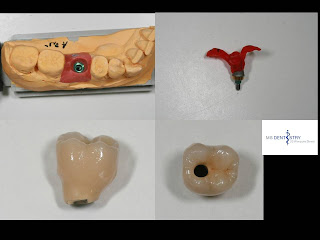
Master model showing the newly regained gum relief.


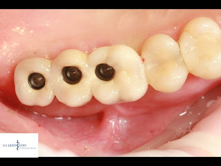
Direct clinical picture showing the nice modelation of the the gum - interdental papilae.


The lab work before seating.


The screw reatined crowns in place before closing the access cavity.


The aesthetic harmony!


X-ray control to check the fit of the rstorations.


Sonntag, 25. April 2010

A combination of cemented and screw reatained way of crown cemebtation


How to avoid failures while seating implant retained crowns?

Incident:

Formation of cement cushion between abutment and crown.

Disadvantage:

Crown can not be seated in full.

How to avoid?

By adequate crown venting - the occlusal acces of the crown will allow evacuation of cement.


Incident:

Abutment screw gets loos over time.

Disadvantage:

Decementation of crown may be difficult to almost impossible resulting in need of destruction.

How to avoid?

Occlusal open crown will allow for future abutment screw tightening.

Samstag, 17. April 2010

Preoperative x-ray showing the failing teeth.
Intraoral picture showing the preoperative clinical situation.

The wound was covered with PRGF once implants have been inserted and bone graft performed.


Control x- ray proving the perfect position of the implants.


Immediate temporaries


Patient received immediate temporaraies once the surgical procedure was completed.